Doppler in Dairy Cattle Reproduction
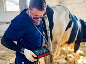
For some time now, ultrasound machines equipped with Doppler functionality have been increasingly used in cattle reproduction. The examination technique itself does not differ from classical rectal ultrasonography—the key difference lies in the presence of Doppler, which enables assessment of blood flow in blood vessels. This gives veterinarians an additional tool to optimize reproduction in dairy herds. When used by an experienced specialist, Doppler provides a significant advantage over standard ultrasound equipment, not to mention traditional manual examination. In field practice, this technology can be successfully used daily by veterinarians involved in animal reproduction.
Lutein cyst on the ovary, wall thickness greater than 3 mm. Lutein tissue present in the wall with Doppler flows.
Doppler facilitates not only more precise pregnancy diagnosis and fetal monitoring but also allows for more accurate assessment of ovarian condition. In clinical practice, and with proper experience, Doppler enables evaluation of uterine blood flow. This information can help determine the stage of the estrous cycle, although interpretation requires some skill. In non-pregnant females, uterine blood flow varies with the cycle phase—closely linked to estrogen and progesterone levels. The highest blood flow occurs during the proestrus and estrus phases, with a decrease in diestrus. Additionally, increased uterine perfusion is observed during the first 21 days of pregnancy.
Pregnancy, cotyledons with Doppler flows.
Even more important is the assessment of follicular perfusion, i.e., the degree of vascularization of ovarian follicles. Scientific studies show that a well-vascularized pre-ovulatory follicle is associated with higher insemination success rates. A dominant or actively developing follicle should exhibit strong blood flow, which also positively influences the hormonal profile. With Doppler, a veterinarian can determine whether a follicle is undergoing atresia and whether perfusion is appropriate. Based on this, one can decide whether the animal is economically and physiologically ready, or whether it’s better to wait until the next heat.
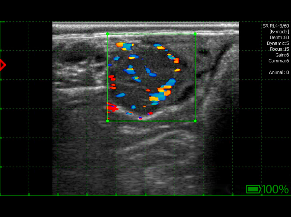
Corpus luteum with normal Doppler flows.
One of the most frequently used applications of Doppler in cattle reproduction is the assessment of corpus luteum (CL) vascularization. The most accurate method for evaluating CL function during pregnancy is progesterone measurement, but this is time-consuming and often economically unjustified. In this context, Doppler becomes a valuable tool for quick and effective assessment of CL activity—useful in both pregnancy diagnosis and estrus synchronization. In pregnancy diagnosis, the biggest challenge is confirming the absence of pregnancy. Doppler allows earlier detection of failed insemination, significantly speeding up the farmer’s response time.
Gynecological examination of the ovary. The image shows a distinct corpus luteum with normal echogenicity and normal Doppler flows. A small follicle is visible next to the corpus luteum.
It’s well known that CL diameter positively correlates with progesterone levels; however, studies have shown that during luteolysis, progesterone levels drop much sooner than CL volume decreases. Doppler eliminates this problem—since the highly vascularized CL loses blood perfusion early in luteolysis, even before shrinking. This enables early detection of failed insemination—even before day 28—which offers a clear advantage over traditional ultrasound. This also makes it possible to resynchronize non-pregnant animals earlier. By incorporating Doppler into routine practice, veterinarians can assess CL function around day 20 post-insemination, identifying CLs undergoing luteolysis.
Gynecological examination of the ovary. The ultrasound image shows two corpora lutea with normal Doppler flows, both corpora lutea have cavities.
Doppler also plays a role in evaluating CLs before animals enter hormonal treatment programs. Blood flow analysis helps assess functional CL activity and predict response to hormonal therapy. This enables precise drug selection, improving reproductive program effectiveness and offering real savings for farmers—faster, more effective synchronization translates to better outcomes.
Gynecological examination of the ovary – three corpora lutea visible with normal Doppler flows.
Doppler is also applied in embryo transfer (ET) as a recipient selection tool. Studies show higher embryo implantation success in cows with well-vascularized CLs compared to those with poor perfusion. Today, CL vascularization is considered the most accurate predictor of ET success—more reliable than CL diameter, surface area, or even progesterone levels.
Gynecological examination of the ovary. Two corpora lutea with Doppler flows and a small follicle are visible.
Doppler technology also shows promise in early synchronization, which can be done as soon as 12–15 days post-insemination. Various hormonal protocols based on progesterone or GnRH are used for this purpose, aiming to shorten intervals between inseminations and increase conception rates. Early synchronization may also be incorporated into ET programs, with appropriate modifications to match the procedure’s specifics.
Gynecological examination of the ovary – corpus luteum with Doppler flows and a small ovarian follicle.
In summary, Doppler ultrasonography in cattle reproduction enables earlier pregnancy detection and more precise evaluation of animals selected for estrous cycle synchronization. This directly improves reproductive efficiency—shortening intervals between inseminations and enhancing key fertility indicators. In the hands of an experienced veterinarian working with larger herds, Doppler becomes a powerful tool for significantly reducing both veterinary and breeding costs. It supports better selection of hormonal treatments, faster elimination of animals with low reproductive potential, more effective insemination candidate selection (reducing semen costs), and earlier pregnancy diagnosis—allowing quicker farmer response.
Lutein cyst – thick wall with Doppler flows.
Furthermore, Doppler improves recipient selection in embryo transfer programs. Beyond the currently known and practiced functions, Doppler’s applications are likely to expand as technology advances and new research emerges. In the future, Doppler is expected to become an essential component of modern reproductive diagnostics and management, helping veterinarians and farmers manage herds with even greater precision and efficiency.

DVM, Michał Barczykowski