Lung ultrasound examination in calves

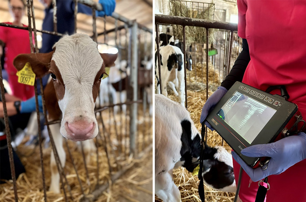
An ultrasound examination in farm animals is often associated with rectal examination. However, this perspective does not fully reflect its applications. Ultrasound is used to diagnose tumours, abscesses, haematomas, and to examine liver, udders, navel and lungs in calves. Lung ultrasound examination in calves is a non-invasive, rapid and objective technique to assess the calf’s breath. Its main advantage is a much higher sensitivity as compared to the use of a traditional phonendoscope. In addition, it allows to determine the extent of lung involvement resulting from possible pathologies. Lung ultrasonography allows confirmation of clinical pneumonia and detection of subclinical pneumonia. A veterinarian specialising in the treatment of farm animals may use this valuable information to support breeders in making more informed decisions about the farm.
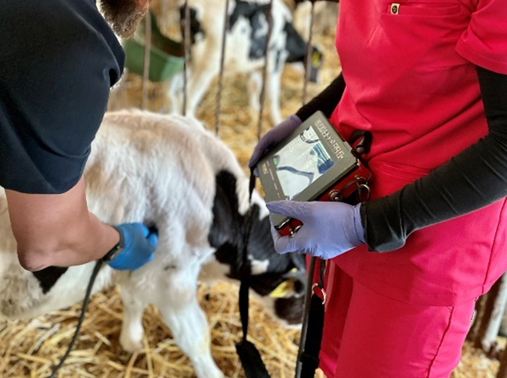
The benefits of this diagnostic method certainly include its simplicity and speed of the examination performance, which, under appropriate conditions, takes only about 60 seconds. A linear probe is used to test the lungs in calves, which is also used in rectal examinations.
The examination technique itself is very simple. Depending on the areas to be visualised, the probe can be applied vertically or horizontally, adjusting it to own preferences. However, in practical experience, it is strongly recommend to focus on the technique of keeping the probe in an upright position. This is when it is possible to accurately visualise the peak parts of the lungs, which are often areas where pathologies are most common. The hardest task in using this method is the identification of the images.
The normal image of the lungs is characterised by the lack of visibility of internal organs due to the accumulated air (the air prevents the transmission of ultrasounds). So, the more advanced the pathology, the more is visible, especially in the case of occurrence of pus, other fluid or fibrotic lesions after previous diseases. In terms of anatomy, cattle lungs constitute an exception because, despite the large size of these animals, their lungs are relatively small. This is why this organ poses the greatest challenge for veterinarians, especially in the case of calves, since it has virtually no ability to regenerate. The authors specialising in bovine ultrasonography suggest to determine own reference points for lung imaging. Such points include the diaphragm, the heart, the pulmonary vessels depending on the left and right lung, and the lobe being examined. The starting pathologies are crucial.
Moreover, the presence of individual artefacts during the examination is an interesting phenomenon. In ultrasound diagnosis, the term “artefacts” refers to the wrong echoes that do not reflect the existing anatomical structures or represent these structures in an incorrect location. Artefacts may be useful for their own purposes, allowing their mutual relationships to be analysed, especially in the context of respiratory system diagnostics. Artefacts in the imaging diagnostics of lung diseases are usually hyperechoic lines. We distinguish the following:
A-line artefacts – horizontal lines along the pleural line. The distance between them is equal to the distance between the pleura line and the probe.
B-line artefacts – vertical lines that originate on the pleural line. They are visible along the entire length of the screen and move in line with the movements of the visceral pleura. They are caused by the presence of a small amount of fluid under the visceral pleura, usually in the interalveolar septa, and usually they do not occur together with A-line artefacts. In one ultrasound scan performed at any site of the chest, one or two B-line artefacts may be expected to be present, which is considered normal.
C-line artefacts – vertical hyperechogenic lines similar to B-line artefacts but beginning at a different site. Their beginning is usually the pathological site which is most interesting. These sites are characterised by increased tissue density than the normal lung tissue.

There are numerous other artefacts, but the three listed above are key from the point of view of diagnostics. When it comes to the limitations of this diagnostic method, it is particularly important that it does not allow imaging of deeper structures. This applies mainly to pathologies that are located in deeper areas of the lungs and are surrounded by air, preventing the penetration of ultrasound radiation. It is best to combine diagnostic methods, e.g. with an ultrasound scan, and perform a conventional auscultatory examination using a phonendoscope. There are also more advanced procedures, such as a lung biopsy. From a practical point of view, this method is not recommended due to high invasiveness, a number of complications and a relatively complicated technique.
In cattle veterinary medicine, non-invasive methods which are both fast and cost-effective are the ones which are most commonly used. In addition to traditional techniques, such as auscultation and percussion of animals and performing bronchoalveolar lavage, ultrasound examinations of the lungs demonstrate these characteristics well.

Vet. Michał Barczykowski


